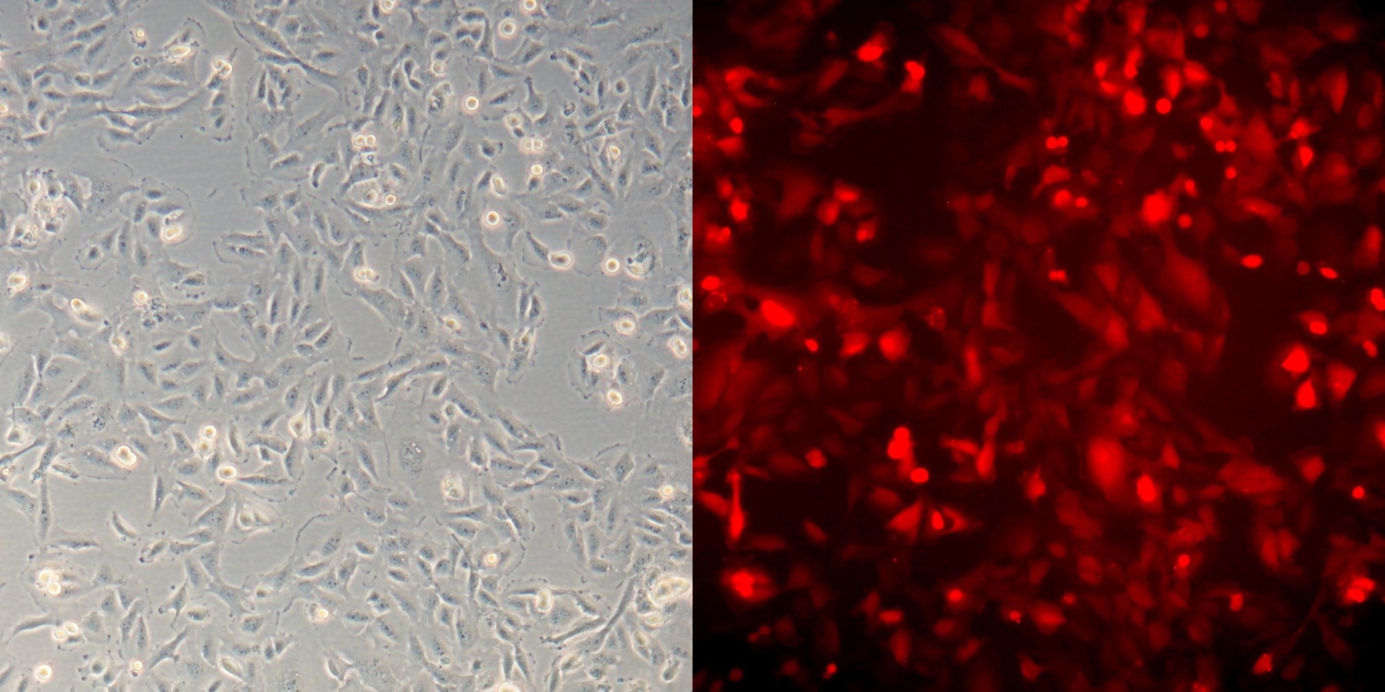
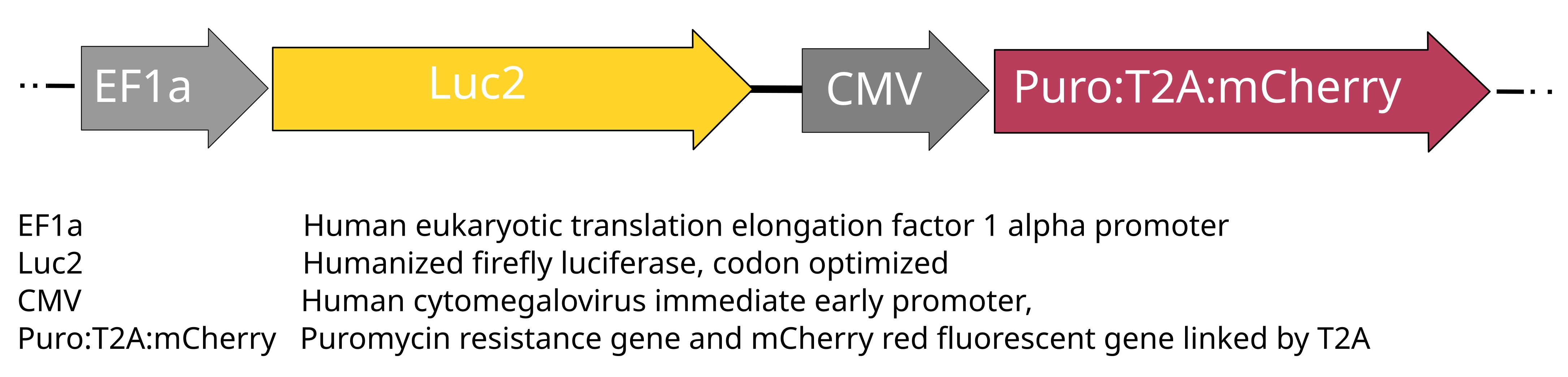
The polyclonal cell line T24-Luc/mCherry (T24-Lch) is derived from the urinary bladder carcinoma cell line T24 and was generated through lentiviral vector-mediated transduction. This vector encodes the codon-optimized firefly luciferase gene (luc2) controlled by the EF-1 alpha promoter, along with the genes for Puromycin N-acetyl-transferase and mCherry red fluorescent protein, linked by a T2A self-cleaving peptide and regulated by the CMV promoter. T24-Luc/mCherry cells demonstrate stable expression of luciferase, mCherry red fluorescence and resistance to Puromycin. The mCherry red fluorescence can be visualized with an excitation at 587 nm and emission at 610 nm.
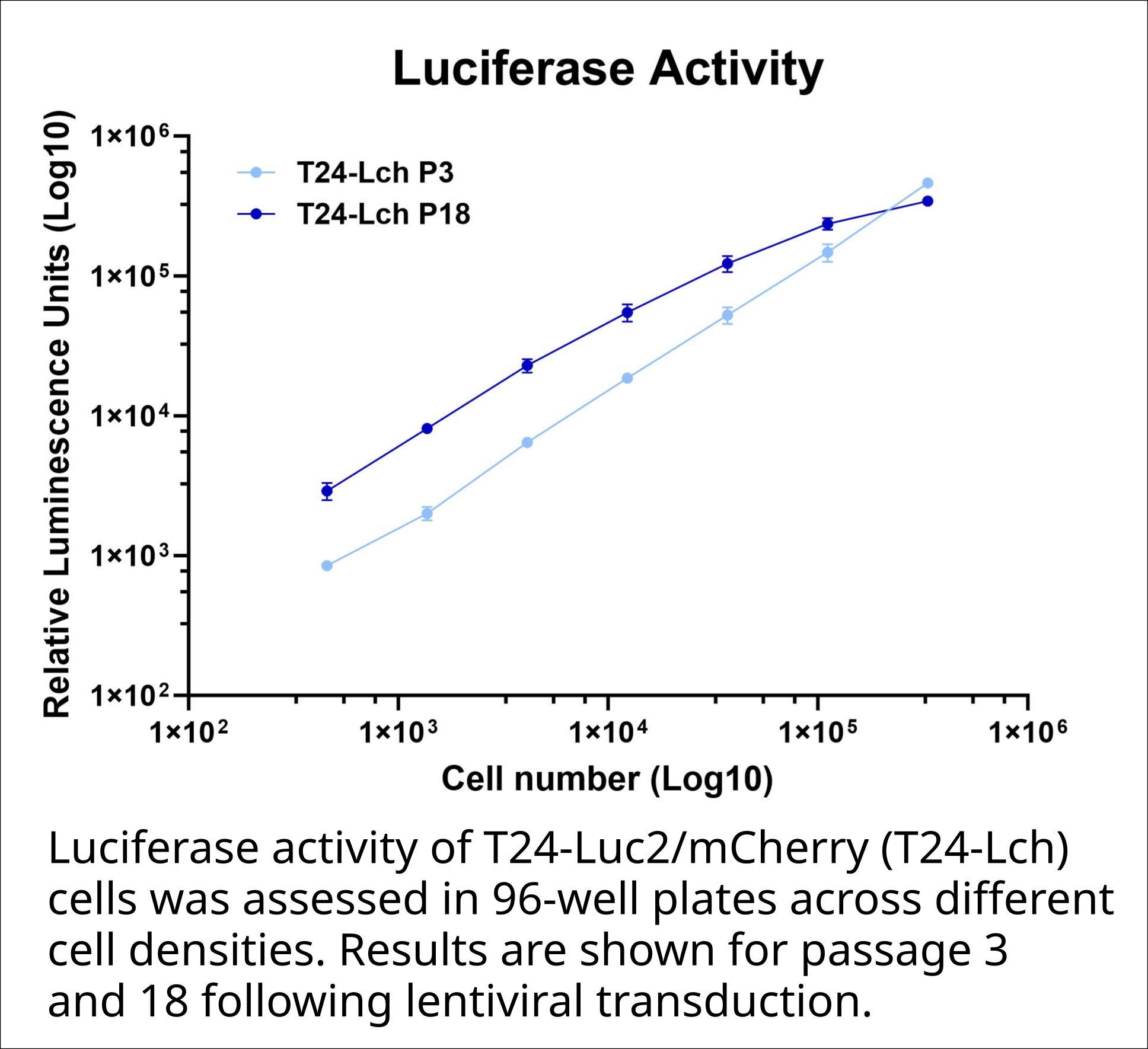
The stable luciferase expression in T24-Luc/mCherry enables highly sensitive and quantitative bioluminescence-based detection of viable cells in-vitro and in-vivo. Luminescence intensity directly correlates with viable cell number, allowing accurate measurement of cell viability, proliferation, and treatment responses. The cell line is particularly well suited for cell-based cytotoxicity assays, including immune effector cell assays such as TDCC, ADCC, and NK cell-mediated killing studies. Furthermore, stable luciferase expression enables sensitive in-vivo bioluminescence imaging, allowing non-invasive longitudinal monitoring of tumor growth, tumor burden, cell persistence, and therapeutic responses in preclinical animal models.
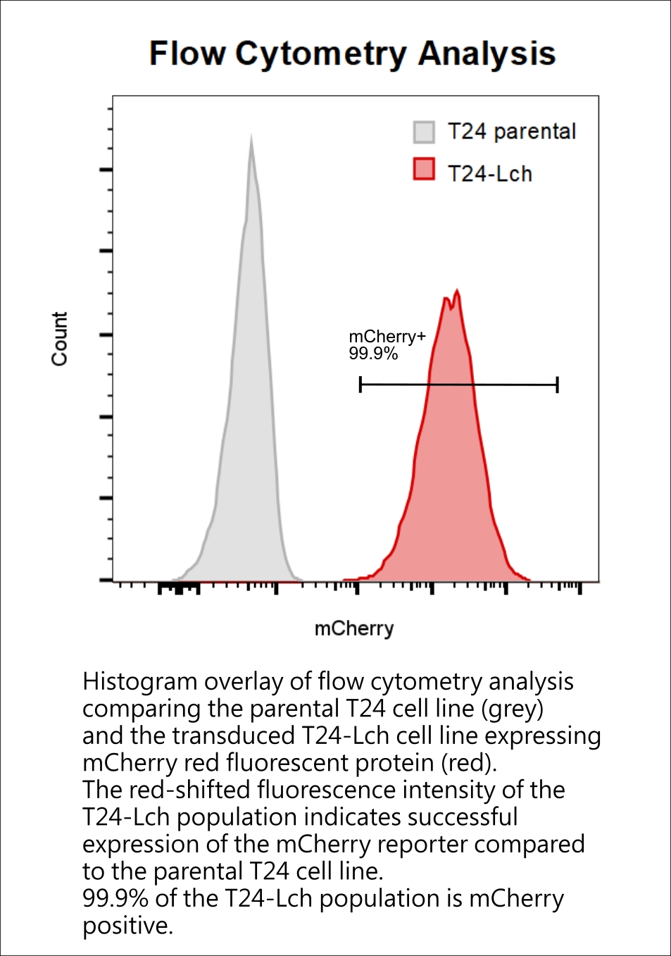
mCherry is a photostable, red fluorescent protein (Ex 587 nm, Em 610 nm) that enables robust visualization of reporter-positive cells by fluorescence microscopy and flow cytometry. Stable mCherry expression allows live-cell imaging without additional staining and facilitates cell tracking in co-culture systems, including T-cell-dependent cellular cytotoxicity (TDCC) and antibody-dependent cellular cytotoxicity (ADCC) assays. Furthermore, mCherry supports flow cytometric identification, isolation, and phenotypic characterization of reporter cells following experimental treatment.
The dual-reporter cell line T24-Luc/mCherry combines highly sensitive quantitative bioluminescence measurements with direct fluorescent visualization of reporter-positive cells. Stable luciferase expression enables accurate assessment of cell viability, proliferation, and treatment responses, while constitutive mCherry expression facilitates cell tracking, identification, and phenotypic analysis by fluorescence microscopy and flow cytometry. Together, the two reporters provide complementary and independent readouts, making the cell line particularly valuable for cytotoxicity assays, immuno-oncology applications, drug screening, co-culture studies, and in vivo tumor monitoring.
Key Features
- Stable luciferase expression enables reliable readout in cell viability and immune cell-mediated cytotoxicity (TDCC, ADCC, NKDCC) assays.
- Luciferase expression supports non-invasive, real-time tumor tracking in in-vivo bioluminescence imaging.
- Constitutive mCherry expression enables direct fluorescent visualization, cell tracking, and flow cytometry-based identification and analysis of reporter-positive cells.
- Stable expression has been verified for at least 15 passages.
- The cell line has been verified to be mycoplasma-free, and cell line authentication has been confirmed.
Applications
- Immune effector cell assays
- T cell or NK cell dependent cytotoxicity (TDCC, NKDCC)
- Antibody dependent cytotoxicity (ADCC)
- CAR-T / CAR-NK assays
- Bispecific antibody testing
- Cell viability & proliferation assays
- Drug cytotoxicity
- Growth inhibition
- Long-term proliferation
- In-vivo Imaging
- Tumor growth monitoring
- Metastasis studies
- Therapy response over time
- Live Cell Visualization
- Cell Tracking in Co-Culture Systems
- Flow Cytometry-Based Identification and Sorting
- Identification of target cells in mixed populations
- Quantification of surviving tumor cells
- Assessment of cell death markers
- Analysis of reporter-cell phenotype after treatment (e.g. apoptosis staining)
Shipping & Storage
- Content: 1 cryovial containing 5 Mio cells in 1ml freezing medium (RPMI1640 with 10% FBS and 5% DMSO)
- Shipping: On dry ice
- Storage: Liquid nitrogen vapor phase
Important:
Store cell cryovials in liquid nitrogen immediately or start culture according to product sheet instructions. Never store cell vials at -80°C as cell viability will decrease.
Cell lines are supplied for research use only and are subject to a Limited Use License as described in the Terms & Conditions. Additional usage rights may be available upon request.